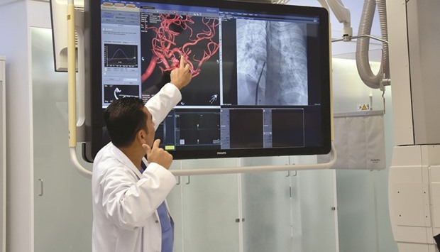
The Hamad Medical Corporation (HMC) has opened a state-of-the-art laboratory capable of producing clearer images that can be used to better diagnose heart, brain and blood vessel problems.
“The lab at the Hamad General Hospital produces high-quality images with lowest radiation, including images from CT scan, MRI, and live echocardiogram. In echocardiogram, it combines both echocardiography and angiography at the same time,” Catheterisation Laboratory Pediatric cardiologist Dr Hesham al-Saloos told reporters last week.
First of its kind in the Mena region, he said the laboratory could handle five to seven patients per day depending on how complex the procedure was. It also has two procedure
rooms: a recovery area and day care.
“If there is a hole in the heart, it can close it without a surgery, and if there is a narrow valve, it can be opened up,” Dr al-Saloos explained. “For vascular, it can open up vessels. For the brain, it can treat stroke in a secluded area. If there is aneurysm, it can be treated
without opening up the brain.”
Some procedures may take three to four hours while other cases only last only about 20 minutes, according to Dr al-Saloos. “If you are doing only diagnostic procedures,
it can do more than 20 patients.”
Out of 1,000 patients seen annually at the hospital, 10% have blocked arteries and another 50 to 100 have aneurysms, according to professor Ashfaq Shuaib, director of Neuroscience Institute at HMC. Aneurysm is an abnormal, blood-filled sac formed by dilation of the wall of a blood vessel or heart ventricle, most commonly the abdominal aorta and intracranial arteries, resulting from disease or trauma to the wall, as in atherosclerosis.
Echoing the statement of Dr al-Saloos, he said the new facility would help them treat patients faster, further improve their capacity and allow them to hire very competent people.
Shuaib noted that patients were normally taken to the emergency for a CT or CAT scan where at least one hour is wasted.
“Every minute, for every artery blocked, a patient is losing 2mn brain cells, so speed is very important,” he pointed out. “Let us say. one hour, because now we are not taking them into the CAT scanner, they are coming here directly, so there are 100mn cells you can protect,” he said.
“What makes it very unique is that in angio, you take pictures but it doesn’t show you much about the function. You are only looking at the arteries but not the brain, this clarity (produced by the lab) gives you a profusion of the brain,” he added.
“So not only does it gives you imaging facilities, it also gives you profusion facilities and you could pull the clot out or put coils in an aneurysm, state-of-the-art, it does not get any better than this.”

An official explaining the functioning of the new machine.
