Researchers at Weill Cornell Medicine-Qatar (WCM-Q) and the Neurosciences Institute at Hamad Medical Corporation (HMC) have won a prestigious international funding for their innovative proposal to use eye examinations to aid in early diagnosis, analysis of disease progression and benefits of treatment in patients with Multiple Sclerosis (MS).
WCM-Q professor of medicine Dr Rayaz Malik was presented with the award for Multiple Sclerosis Innovation by the European Committee for Treatment and Research in Multiple Sclerosis. It was one of only four research grants awarded from a total of 260 applications from 45 countries and the first ever to be awarded to the Mena region.
The research team will now use the grant to conduct a comprehensive 24-month study to determine whether Corneal Confocal Microscopy can be a viable method for determining nerve damage in patients with MS. The research has local significance as Qatar appears to have a higher than expected prevalence of MS.
MS is a debilitating disease of the central nervous system in which nerve impulses within the brain, and between the brain and other parts of the body, are disrupted. Symptoms include difficulty walking, vision problems, fatigue, pain and cognitive changes.
MS is difficult to monitor as each patient is affected in different ways and experience different rates of disease progression. Additionally, the most widely used monitoring technique, magnetic resonance imaging scans of the brain, cannot accurately identify nerve damage, which is the underlying pathology associated with progressive neurological deficits.
The new test is non-invasive and utilises existing ophthalmic equipment that many hospitals already have.
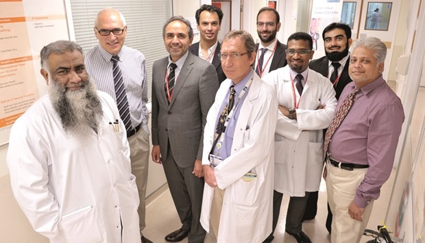
Dr Malik and his team of other WCM-Q researchers and neurologists from HMC, Dr Saadat Kamran, senior consultant neurologist, and Dr Ashfaq Shuaib, professor of medicine and neurology and director, Neuroscience Institute, believe their technique could provide a more accurate and easier method for monitoring MS.
The cornea has the densest concentration of nerve fibres anywhere in the body. Dr Malik and colleagues have pioneered the technique of ‘Corneal Confocal Microscopy’ over the last 15 years to enable close examination and imaging of the cornea’s nerve fibres to identify nerve damage in a variety of conditions including diabetic neuropathy, hereditary neuropathies and in patients with Parkinson’s disease.
Dr Malik said: “MS is an extremely distressing disease, which is very difficult to monitor and has limited treatment options. There is an urgent need for a new monitoring tool that is both reliable and more accurate that could be applied both in the clinic and in clinical trials. Our research indicates that nerve damage in the cornea is a reliable indicator of nerve damage in the brain that characterises MS.”
Dr Ashfaq Shuaib of HMC said, “We are extremely pleased to be involved in this important research and this award gives us all a great opportunity to continue to work together to develop a very effective new tool for diagnosing and assessing progression of MS.”

Researchers from WCM-Q and HMC who are working together to develop an innovative monitoring tool for MS.
