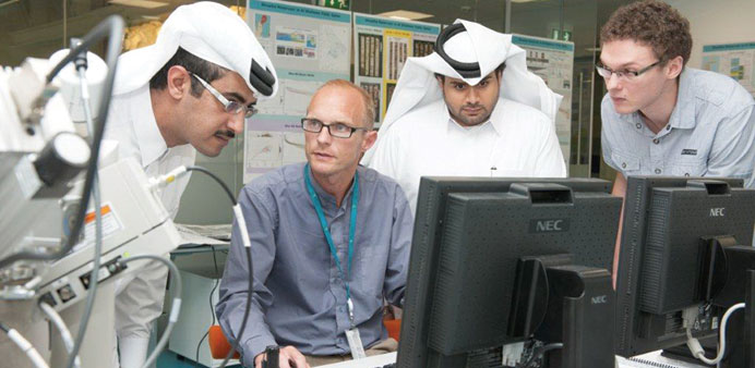
As the Qatar Museums Authority celebrates the unveiling of the country’s first Unesco World Heritage listed site tomorrow at Al Zubarah, a team of researchers at the Qatar Science and Technology Park are proud of their small role in uncovering the country’s history.
The researchers, based at the Maersk Oil Research and Technology Centre (MO-RTC), use the latest cutting-edge technology to scan rock samples at a micro level and create images that assist in oil recovery, and now archaeology.
The MO-RTC Digital Core Laboratory is the first of its kind to operate in the Middle East and is home to the region’s only electron microscope with associated QEMscan capability.
Using the techniques of computed tomography (CT) and quantitative automated mineralogy, archaeologists can compare the mineralogy of pottery sherds with that of locally sourced clays, and understand the origins of the ceramics from sites under excavation within Qatar.
Abdulrahman al-Emadi, head of MO-RTC, said research from the Digital Core Laboratory can help improve oil recovery rates but – through its role in archaeology – it’s also a fantastic example of industry and academia working together for the greater good of Qatar.
The CT and quantitative automated mineralogy techniques at the MO-RTC have helped researchers discover important information on the far-reaching trade routes of the Islamic period that extended to regions far beyond the Arabian Gulf.
Evidence for this trade comes from pottery on several Islamic sites within Qatar, including the town of Al Zubarah on the north-west coast.
Theis Ivan Solling, the manager of Maersk Oil’s Digital Core Laboratory, explained how the procedure is carried out: “Small sections are cut from each sherd using a diamond-bladed saw, placed in a mould, coated in epoxy resin and left to solidify for 24 hours. A thin slice is then cut from the hardened plug. It is then glued to a microscope slide and polished to an ultra-thin state. We then do experiments to map out the mineralogy in two dimensions. The section is subjected to an electron beam. The data is then treated to yield information about the mineralogy. Geologists can then identify which part of the world that particular mineral came from. It’s like a fingerprint, each is unique.”
In another part of the lab, CT scanning produces three-dimensional images of the structure of a pottery sample.
“A total of 13,000 images are used to make a mathematical reconstruction. This results in a file that records the spatial structure of the specimen. From the results researchers can see how all the pores are interconnected, and this yields information not only about the production method of the sherd but also its origin,” Solling added.

A group of researchers at work at the Maersk Oil Research and Technology Centre.
